

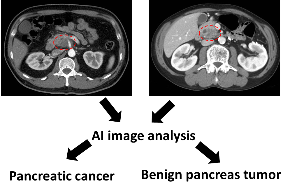
Pancreatic cancer (PC) is the most lethal cancer, and its incidence and mortality rates are rising. Computed tomography (CT) and magnetic resonance imaging (MRI) are the major tools for detection and evaluation of pancreatic masses. However, CT and MRI have only modest accuracy in determining the nature of the masses (malignant vs non-malignant), and approximately 40% of pancreatic tumors <2 cm are missed. Furthermore, the interpretation of CT and MRI is based on subjective experiences with considerable interobserver variations. Therefore, 85% of PCs are diagnosed at a late stage and not amenable to surgical resection, underscoring an urgent need for novel methods to improve the diagnostic ability of CT and MRI for pancreatic masses.
The major aim of this project is development and commercial release of an artificial intelligence (AI)-assisted image analysis software that enables early detection and accurate diagnosis of pancreatic masses by CT/MRI. This software aims to detect CT/MRI features that are undetectable by human eyes to enable detection of small pancreatic masses, accurately differentiate between cancerous and non-cancerous masses, and improve its accuracy by deep learning. In this 4 year project, we aim to develop the alpha version, beta version, and release candidate of the software by the end of year 2, 3, and 4, respectively. In the first and second year, we will establish an annotated database of pancreatic CT/MRI images and analyze those images using radiomics, statistical shape analysis, and deep learning. In the third year, the alpha version will be tested in real-world clinical practice at National Taiwan University Hospital to improve its ability in detecting/classifying pancreatic masses, and to refine the user interface. The resultant beta version will undergo prospective multiphase multicenter clinical testing and fine-tuning in the fourth year to produce the release candidate. We will apply for software patent and seek commercial release of the software. In addition, we plan to publish 4 research papers on the performance of the software in comparison with human interpretation of CT/MRI and invasive pancreatic tissue acquisition.
The other major aim of this project is to establish an interdisciplinary/international network and cultivate young talents for the AI sector of Taiwan. By bringing together medicine, mathematics/statistics, computer science, and business, this network will benefit researchers and industry and serve as a platform for advancing the application of AI in medicine.
Accurate detection and diagnosis of pancreatic masses is a challenge that doctors face every day, and misdiagnosis often leads to grave consequences. If our aims are achieved, our software may solve these problems and significantly transform/improve clinical care, saving lives and costs. Therefore, the software has significant academic, clinical, and industrial values. Furthermore, this project can serve as a proof-of-concept that AI can conduct data mining on the vast amount of information in CT and MRI to improve the diagnostic ability. The experience and methodologies that are generated from this project can be applied to many other unsolved clinical problems.
◎ PI

PI Wei-Chih Liao
Associate Professor, Department of Internal Medicine, College of Medicine, NTU

Co-PI Kao-Lang Liu
Attending Radiologist, Body Imaging Section, Department of Medical Imaging, NTUH

Co-PI Weichung Wang
Professor, Graduate Institute of Applied Mathematical Sciences, NTU